What is a vitrectomy?
 Vitrectomy is a type of eye surgery that treats disorders of the retina and vitreous.
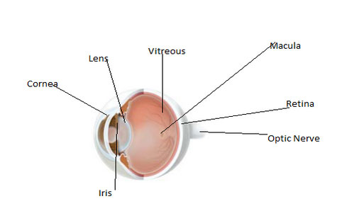
The retina is the light-sensing tissue at the back of the eye. The vitreous is the clear, jelly-like substance that fills the middle of the eye. The vitreous is removed during vitrectomy surgery and usually replaced by a saltwater solution.
When do you need a vitrectomy?
Your ophthalmologist (Eye M.D.) may recommend vitrectomy surgery to treat the following eye problems:
- Diabetic retinopathy, if bleeding and scar tissue is present;
- Some retinal detachments;
- Infection inside the eye;
- Severe eye injury;
- Macular pucker (wrinkling of the retina);
- Macular hole (partial loss of vision for fine details);
- Certain problems after cataract surgery.
How can a vitrectomy improve vision?
Vitrectomy surgery often improves or stabilizes vision. The operation removes any blood or debris (from infection or inflammation) that may be blocking or blurring light as it focuses on the retina.
Vitrectomy surgery removes scar tissue that can displace, wrinkle, or tear the retina. Vision is poor if the retina is not in its normal position.
This surgery can also remove a foreign object stuck inside the eye as the result of an injury. Most foreign objects will damage vision if they are not removed.
What happens if you decide to have a vitrectomy surgery?
before surgery: Your ophthalmologist will decide whether local or general anesthesia is best for you. You may have to stay overnight in the hospital. Before surgery you will need to have a physical examination to alert your ophthalmologist to any special medical risks. A painless ultrasound test may be performed before the surgery to view the inside of the eye.
vitrectomy surgery: The length of the operation varies from one to several hours, depending on your condition. In certain situations, your ophthalmologist may do another surgical procedure at the same time, such as repairing a detached retina or removing a cataract.
Your ophthalmologist performs the operation while looking into your eye with a microscope.
Various miniature instruments are placed into the eye through tiny incisions in the sclera (white part of the eye).
Vitrectomy surgery removes vitreous clouded with blood that might be causing blurry vision.
In order to get the best possible vision for you, your ophthalmologist will do one or more of the following:
- Remove all cloudy vitreous;
- Remove any scar tissue present, attempting to return the retina to its normal position;
- Remove any foreign object that might be in the eye;
- Treat the eye with a laser to reduce future bleeding or to fix a tear in the retina;
- Place an air or gas bubble in the eye to help the retina remain in its proper position (the bubble will slowly disappear on its own);
- Place silicone oil in the eye, which usuallyrequires later surgical removal.